A Novel Insight into Pharmaceutical Polycrystalline Powders
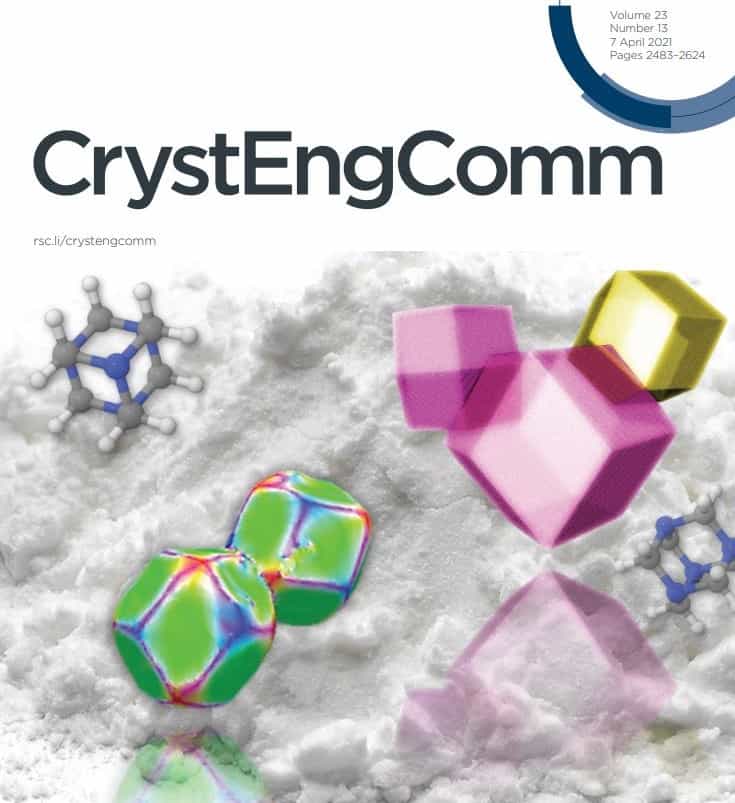
A recent publication by Seda’s Parmesh Gajjar with The University of Manchester featuring on the front cover of the Royal Society of Chemistry Journal CrystEngComm revealed how individual crystals interact within an organic powder bed. In this latest insight, Parmesh shares the results from the publication ‘Crystallographic tomography and molecular modelling of structured organic polycrystalline powders’, and why these results are important.
Understanding polycrystalline powders is vital for informing and improving drug product development, since the interactions between different crystals can play a pivotal role in the behaviour of a formulation. For example, in a dry powder inhaler formulation used for treating respiratory conditions such as asthma and COPD, the interaction between drug crystals and excipient crystals is key to the deagglomeration that allows the formulation to be inhaled directly into the lung. Crystal-crystal interactions also play a role in powder agglomeration, which can affect powder flowability and thus affect many manufacturing processes. Despite this importance, measuring crystal interactions within pharmaceutical powder beds is extremely challenging.
To understand the crystallographic reasons behind powder bed structure, it is important to understand the morphology of crystals in 3D within a powder bed and see how crystal orientations drive crystal-crystal interactions. Using a combination of attenuation X-ray computed tomography (CT, a non-destructive X-ray microscopy technique capable of 3D imaging of particle morphology and powder micromeritic properties) and diffraction contrast tomography (DCT, a diffraction tomography technique that has evolved to enable 3D mapping of crystalline structures) in combination with molecular modelling (modelling based on molecular interactions), it was possible to answer these questions for the organic compound hexamine.
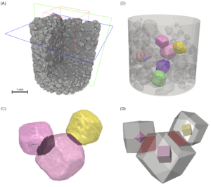
As shown in the figure below, the tomography techniques give unique 3D insight into the structure of the powder bed, and how several crystals cluster together in agglomerates.

Novel imaging methods allow a 3D view of the bed (A), orientation of crystals (B) as well as specific interactions between clusters of crystals (C), with modelling techniques allowing these interactions to be recreated (D).
Image modified from Gajjar et al (2021) with permission under a creative commons license.
Furthermore, the rhombic dodecahedral morphology of crystals could be seen for the first time in 3D within the powder bed alongside exactly how individual crystals interact with each other. Not only were these imaging results the first application of the DCT technique to organic materials, but they were also a powerful compliment to the molecular modelling which provided the interaction energies for different types of crystal interactions. The face-to-face crystal interactions that were experimentally observed were the most energetically preferential. Together, all of these results provide new insight for different mechanisms of powder bed agglomeration. The work certainly shows great potential for unlocking the behaviour of many other pharmaceutical polycrystalline powders, and the next step is to apply the techniques to real formulations, such as dry powder inhaler formulations, to examine how drug and excipient crystals both interact with each other.
This work clearly highlights the important role solid state knowledge plays in the formulation process, and Seda has a depth of expertise in both solid state analysis and pharmaceutical compound behaviour (Talbir Austin). Furthermore, the work shows how imaging techniques provide valuable insight into the different stages of formulation development, and Seda scientists have experience in imaging of formulation components and design (Parmesh Gajjar, Joanna Denbigh) through to imaging of formulations ex-vivo (Claire Patterson).
